Symptoms or appearance
There should be a meaningful reason to treat. An incidental nodule without symptoms often needs monitoring rather than ablation.
 Call the practice
Understand what a thyroid nodule is, what a Bethesda result means, and how ultrasound-guided microwave ablation may be considered for selected benign nodules.
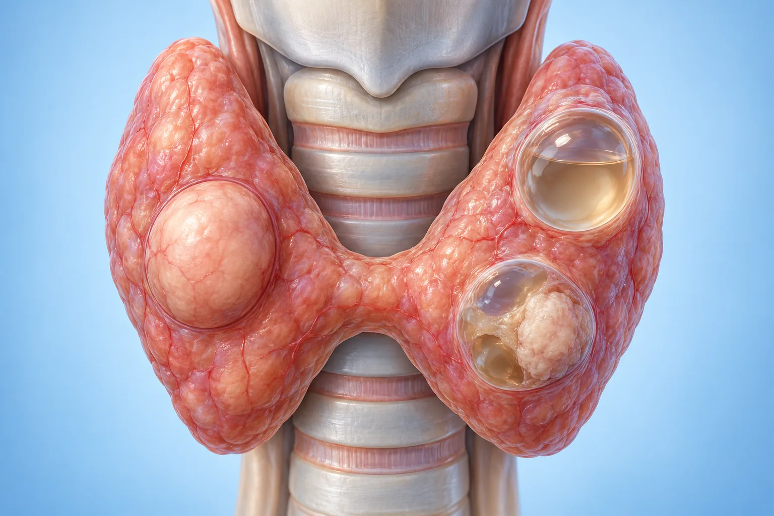
The medical illustrations are AI-generated for education. They do not depict an actual patient or show exact equipment or technique; individual anatomy and care vary.
The thyroid is a small, butterfly-shaped gland at the front of the neck. A nodule is a distinct lump within it. Nodules are common, and many are benign. Most do not cause symptoms and do not need a procedure.
Nodules can be solid, fluid-filled (cystic), or a mixture of both. Some are single; others occur in a gland with several nodules. A nodule’s appearance on ultrasound, growth, symptoms, thyroid blood tests and needle-sample result are considered together.
Bethesda is a six-part system used by the cytology laboratory to describe cells collected from a thyroid nodule by fine-needle aspiration (FNA). It helps guide the next step, but it is interpreted alongside ultrasound and the clinical picture.
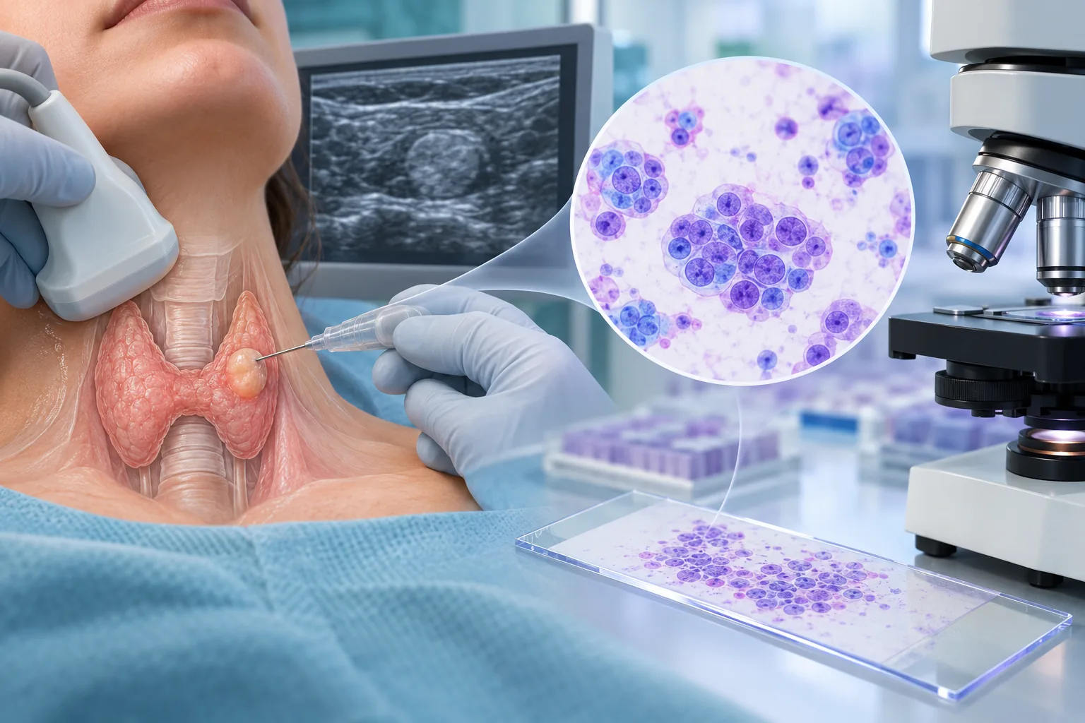
Category names follow the 2023 Bethesda System for Reporting Thyroid Cytopathology. Risk estimates vary by setting and are not used here as a personal prediction.
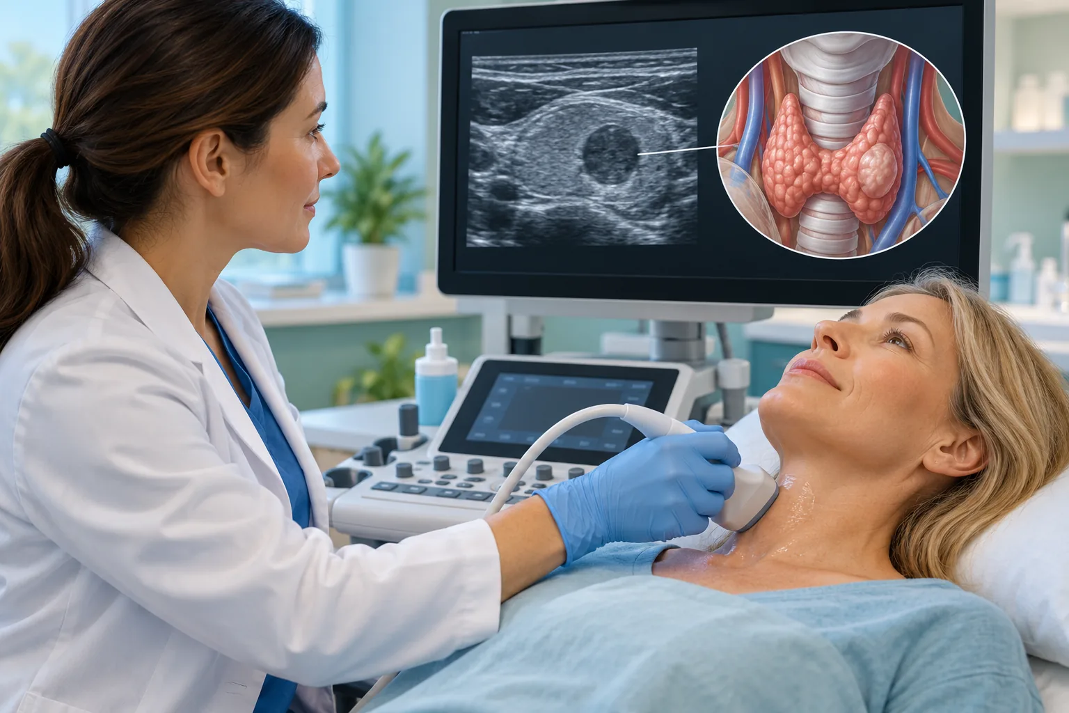
Microwave ablation (MWA) may be discussed when a nodule is confirmed as benign and is causing troublesome pressure, swallowing symptoms, or a visible cosmetic concern. Some people prefer to avoid or defer surgery after discussing the advantages, limitations and alternatives.
Assessment commonly includes a neck ultrasound, thyroid function blood tests and one or more needle samples. For solid or mixed nodules, the 2026 ISVIR consensus recommends two benign FNA results (Bethesda II) in most cases. One benign sample may be sufficient for classic benign ultrasound patterns, such as spongiform appearance or comet-tail artifact. The treating team decides what is appropriate for the individual nodule.
There should be a meaningful reason to treat. An incidental nodule without symptoms often needs monitoring rather than ablation.
The team checks the cytology report against the ultrasound and may request repeat sampling if results are uncertain or discordant.
Location, size, nearby structures, general health and patient preference all matter. No single feature guarantees eligibility.
The Indian Society of Vascular and Interventional Radiology (ISVIR) consensus describes a complete care pathway. It was developed through a modified Delphi process; its 19 statements ultimately reached more than 85% expert agreement. Agreement between experts is not a guarantee of treatment benefit for an individual. These steps translate the recommendations into patient language. Your own plan depends on your scan, sample results, health and preferences.
Ablation is for selected people, not every nodule. For a small nodule under 2 cm, the consensus allows case-by-case consideration when it causes pressure symptoms, a cosmetic concern or significant anxiety; this is a conditional recommendation based on low-level evidence, not a rule to treat. For nodules over 2 cm, it identifies low or very-low-suspicion ultrasound patterns (ACR TI-RADS 1 to 3), or selected intermediate-suspicion patterns (TI-RADS 4) when cytology is benign and symptoms or cosmetic concerns are present. Your specialist interprets the scan category; patients should not try to assign it themselves. Size alone does not mean treatment is needed.
The specialist reviews the ultrasound risk pattern and needle-sample result together. For solid or mixed nodules, the ISVIR consensus recommends two benign fine-needle samples (Bethesda II) in most cases. One benign sample may be enough when ultrasound shows a classic benign pattern, such as a spongiform nodule or comet-tail artifact. The clinician decides whether repeat sampling is appropriate. Suspicious or malignant results need a different diagnostic pathway.
The consensus recommends a complete blood count, a blood-clotting panel and thyroid-function tests (TSH, T3 and T4). If TSH is low, the clinician may arrange a thyroid scan to see whether the nodule is overactive. Overactive nodules are a separate decision pathway; the consensus says ablation may be considered in selected cases but not for multiple autonomous nodules. Other tests depend on the history and findings; calcitonin and thyroglobulin are not recommended routinely for every benign nodule. Tell the team about blood thinners and antiplatelet medicines. Never stop or change them yourself; any adjustment must be planned with the prescribing clinician.
MWA is a thermal treatment and is most often considered for solid or mostly solid nodules, including selected spongiform nodules. For a mainly fluid-filled (cystic or colloid) nodule, the consensus identifies ethanol treatment as a common first option; thermal ablation may be considered in selected cases if there is a substantial solid part or symptoms return. The consensus does not identify microwave as universally superior to other thermal methods; the choice depends on nodule features, the treating team’s experience and available equipment. This guide explains MWA only.
The thyroid lies close to the windpipe, swallowing tube, major blood vessels and nerves that affect the voice. If a nodule is less than 5 mm from important structures, the consensus recommends hydrodissection: the clinician injects fluid under ultrasound to create a temporary protective space. Whether this is needed is decided from your own scan.
MWA is performed with ultrasound guidance and local anaesthetic according to the care plan. The consensus favors treating the nodule in small planned zones by repositioning the antenna under ultrasound (the moving-shot approach). Temporary neck swelling is expected and should be monitored. The team will advise on suitable pain relief and when to seek help.
The consensus recommends clinical review, ultrasound and thyroid-function tests at 1, 6, 12, 24 and 36 months. It defines treatment success as more than 50% reduction in nodule volume at 6 to 12 months; this is a consensus measure, not a promise of symptom relief or an individual outcome. Larger nodules over 20 mL may need more than one session; the consensus says an early repeat may be considered within 6 months. Another session may also be discussed if shrinkage is less than expected, symptoms or cosmetic concerns remain, or the nodule regrows. Timing is individualized by the treating team.
The right choice depends on the diagnosis, symptoms, ultrasound appearance and your priorities. A clinician should explain the options without assuming that a procedure is necessary.
For many benign nodules, follow-up and repeat ultrasound are appropriate.
A needle-like antenna heats targeted tissue through the skin under ultrasound guidance.
May be preferred for some nodules, uncertain diagnoses, or when other clinical factors support it.
Discuss expected benefits, risks, alternatives, aftercare and what matters most to you.

MWA is an image-guided treatment. A thin antenna is placed through the skin into the selected nodule while the clinician watches its position on ultrasound. Microwave energy heats the targeted tissue. The treated tissue is gradually absorbed by the body, so the nodule may shrink over time rather than disappearing immediately.
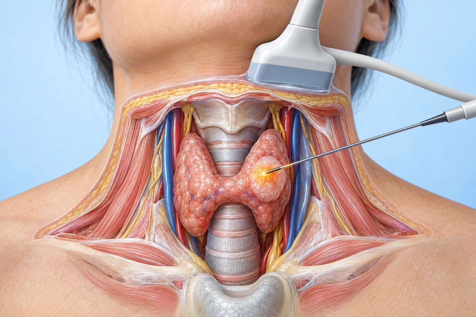
The care team confirms the plan, checks relevant test results and explains consent. The procedure is commonly performed with local anaesthetic, with additional comfort measures depending on the service. You lie with your neck supported and extended.
Under ultrasound, the clinician guides the antenna into the nodule and treats targeted areas. The position may be adjusted to cover the planned tissue while protecting nearby structures. The ISVIR consensus favors treating the nodule in small planned zones by repositioning the antenna under ultrasound (the moving-shot approach). For spongiform nodules, it recommends careful monitoring of heat spread. If the nodule lies within 5 mm of important structures, the consensus recommends hydrodissection, where the clinician uses fluid under ultrasound to create a temporary protective separation. The exact method, duration and monitoring vary by nodule and centre.
You are observed afterward and given instructions for home. Ask who to contact if you develop symptoms after leaving.
Review ultrasound, sample results, symptoms and treatment goals.
Clean the skin, use local anaesthetic as planned, and support the neck.
Place the antenna into the target under real-time imaging.
Apply energy to planned areas; monitor you and provide aftercare advice.

The ISVIR consensus recommends blood-count, clotting and thyroid-function checks before ablation. A low TSH may lead to a thyroid scan; other investigations depend on your history and findings. Preparation differs between centres, so follow your clinic’s instructions. Bring your ultrasound and biopsy reports if requested.
For a suitable benign nodule, MWA aims to reduce its volume and ease related pressure or cosmetic concerns while avoiding an operation and a neck incision. Improvement and shrinkage are gradual and vary. Some patients need continued monitoring, repeat treatment, or another option. The ISVIR consensus notes that nodules larger than 20 mL may need multiple sessions; repeat treatment can also be considered if regrowth, persistent symptoms or inadequate shrinkage occurs.
Possible problems include pain, tenderness, bruising and temporary swelling. Less common but important complications include bleeding, skin injury, voice change from nerve irritation or injury, and damage to nearby structures. The consensus also discusses rare nerve complications, thyroid-function changes and nodule rupture. Its pooled complication figures cover different techniques and patient groups, so they cannot predict your personal risk; your clinician can explain the risks for your case.
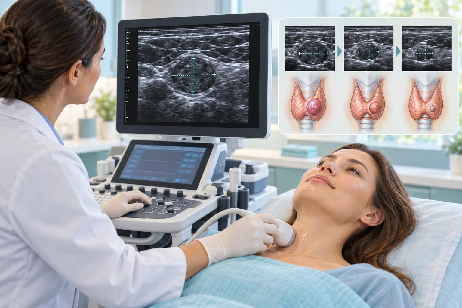
Many people go home the same day after observation, but the plan depends on the centre and individual circumstances. Mild soreness, bruising or temporary swelling may occur; the ISVIR consensus says swelling is expected and should be monitored. Ask when to return to usual activities and what pain relief is suitable for you.
Follow your written instructions. Monitor the neck and contact the care team if symptoms worsen or concern you.
Nodule shrinkage is gradual. The team may review symptoms, ultrasound measurements and thyroid tests when indicated.
The ISVIR consensus recommends clinical review, ultrasound and thyroid-function tests at 1, 6, 12, 24 and 36 months. It defines treatment success as more than 50% volume reduction at 6 to 12 months; this is a consensus measure, not a promise of symptom relief. Keep follow-up appointments even if you feel well, and tell the clinician if the lump grows or symptoms return.
No. Bethesda II describes a benign cytology result. If a nodule causes no important symptoms, monitoring may be enough. Ablation is a choice for selected cases after the diagnosis and options are reviewed.
Usually not. The aim is for treated tissue to shrink gradually. The amount and timing vary, and follow-up helps assess the response.
This guide is about selected benign nodules. A suspicious or malignant result needs a specialist cancer-care pathway. Do not use this page to decide on treatment for a cancer diagnosis.
No. They use different energy sources. This page focuses on microwave ablation only. Evidence and recommendations for one technique should not automatically be assumed to apply to another.
Some nodules do not shrink as much as expected, or can grow again. Further monitoring, another treatment session or a different option may be discussed based on your follow-up.
Many people retain thyroid function, but individual outcomes differ and thyroid blood tests may be followed when appropriate. Ask what monitoring is recommended for you.
The guide uses a focused narrative search of PubMed-indexed clinical studies and systematic reviews on microwave ablation (MWA) of benign thyroid nodules, alongside direct MWA-versus-radiofrequency (RFA) comparisons. It prioritizes 2026–2017 evidence and consensus statements. The 2026 ISVIR Indian consensus and 2026 Indian Association of Endocrine Surgeons consensus are included as professional guidance; the 2026 international thermal-ablation consensus, ATA statement and RFA reporting consensus are also identified by scope. RFA-only findings are not presented as proof of MWA-specific results. This is a focused narrative source review, not a registered systematic review.
Bring your ultrasound and cytology reports if available. The appropriate next step depends on your individual assessment.
Phone: +91 70348-39446 · Email: veindoctoraju@gmail.com