Medical care first
Often the initial approach. The exact plan depends on symptoms, pregnancy goals, other health factors and response to treatment.

If adenomyosis symptoms continue despite conservative treatment and hysterectomy has been discussed, uterine artery embolisation (UAE) may be an option to ask a gynaecologist and interventional radiologist about. It is not first-line care, is not suitable for everyone, and cannot guarantee that hysterectomy will be avoided.
See when UAE may be discussed ↓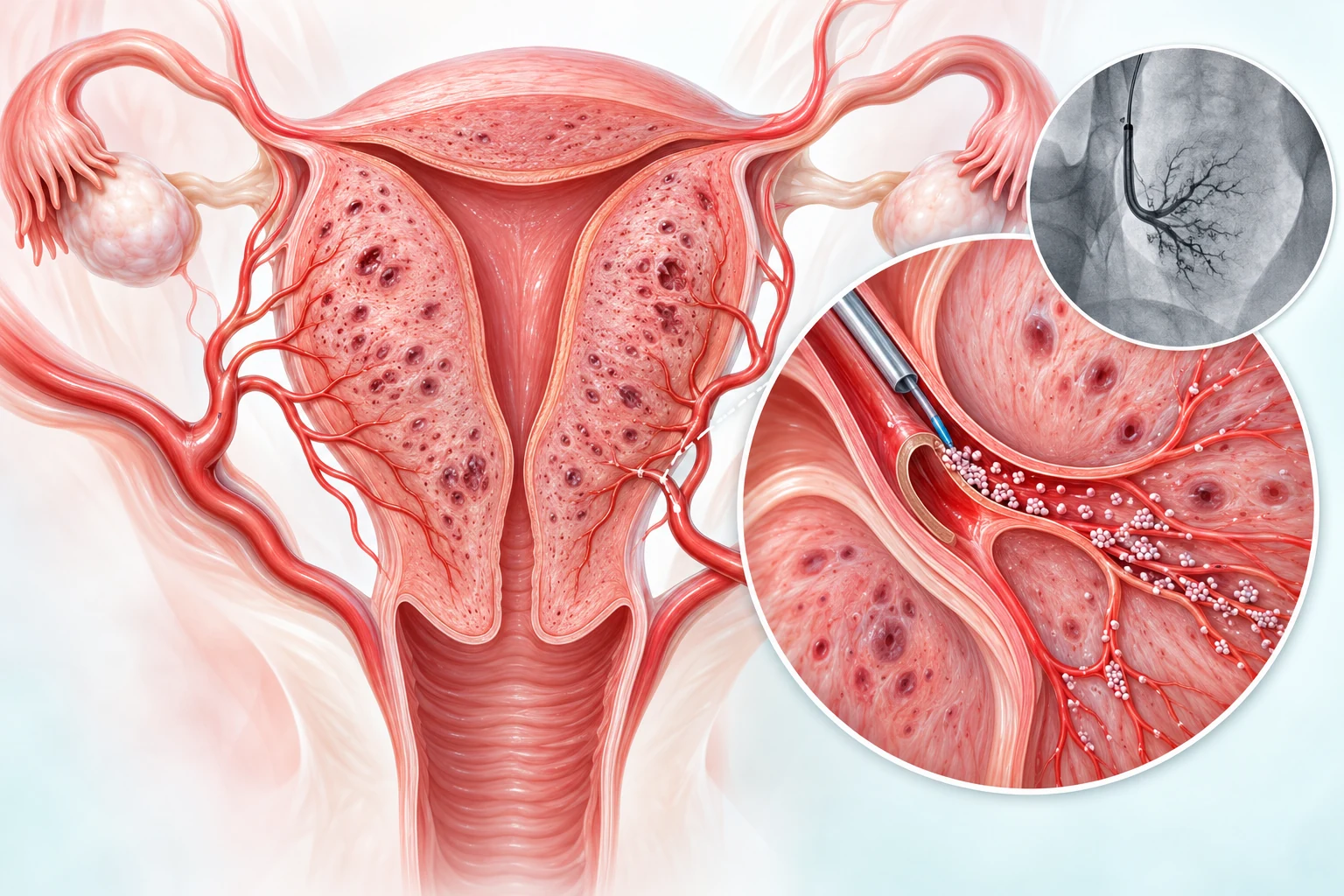
Adenomyosis is a benign condition in which tissue resembling the lining of the uterus is found within its muscular wall. The muscle can become thickened and the uterus may enlarge. It may be diffuse, affecting a broader area, or focal, forming a localized area sometimes called an adenomyoma. Adenomyosis is different from fibroids, although both can occur together.
Some people have no symptoms. Others may experience heavy or prolonged menstrual bleeding, painful periods, pelvic pain or pressure. These symptoms can have other causes too, so assessment matters.
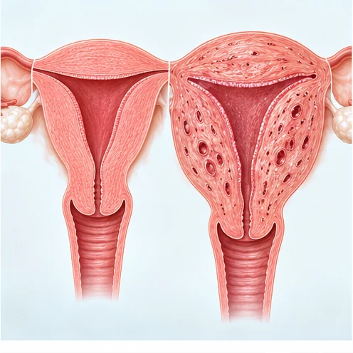
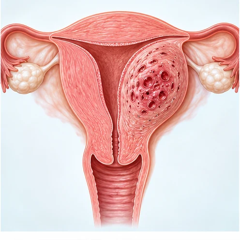
Assessment usually includes a review of bleeding and pain, medical history, examination as appropriate, and pelvic imaging. Transvaginal ultrasound is commonly used; MRI may help clarify the pattern, extent, or other pelvic conditions. Imaging findings should be interpreted with symptoms and clinical context, because no single scan measurement answers every question.
Your team may also evaluate other reasons for bleeding or pain, including fibroids, endometriosis, endometrial problems or infection, when relevant.
Medical options are commonly considered first and chosen around symptoms, health history and preferences. These may include pain-relieving medicines, medicines to reduce bleeding, hormonal treatments such as a levonorgestrel intrauterine system or progestins, and other clinician-directed options. A gynaecologist can explain what is appropriate and the likely benefits and trade-offs.
Often the initial approach. The exact plan depends on symptoms, pregnancy goals, other health factors and response to treatment.
May be discussed after medical care has not helped, is unsuitable, or is not tolerated—particularly when hysterectomy is being considered and the patient wants to explore a uterine-preserving option.
Removing the uterus ends uterine adenomyosis, but it is major surgery and permanently ends the ability to carry a pregnancy. The surgical and recovery considerations should be discussed with the gynaecologist.
A discussion may be reasonable for a person with troublesome symptoms and imaging consistent with adenomyosis, when conservative treatment has not provided enough relief, and when hysterectomy is being considered but the person wants to understand uterus-preserving options. The gynaecologist and interventional radiologist should review the case together where possible.
Whether UAE is appropriate depends on the diagnosis, pattern of disease, symptoms, other pelvic conditions, overall health and personal priorities. A specialist may advise against it or recommend another option.

The interventional radiologist uses X-ray imaging to guide a fine catheter from an artery in the wrist or groin to the uterine arteries. Small particles are delivered to reduce blood flow to the uterine tissue affected by adenomyosis. The access point is small; the procedure still requires careful planning, pain control, monitoring and follow-up.
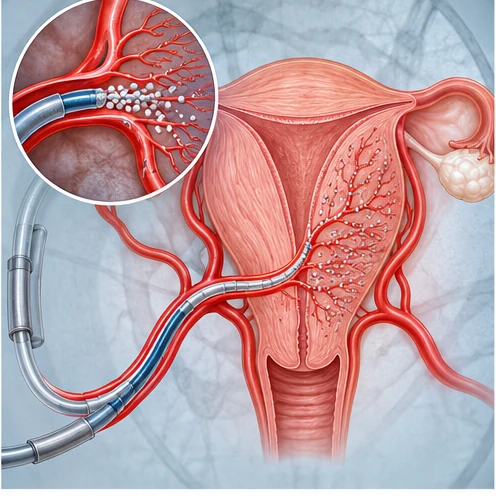
The team reviews symptoms, prior treatment, ultrasound/MRI, goals, medicines and relevant tests.
You discuss expected benefits, limitations, alternatives, risks, pain-control and the possibility of later surgery.
A catheter is guided under X-ray and particles are delivered through the uterine arteries, if the procedure proceeds.
The team treats post-procedure pain, checks recovery and arranges review of symptoms and imaging when appropriate.
Published studies report that some patients experience less bleeding and pain after UAE, but much of the evidence comes from observational cohorts with different patient groups and follow-up periods. Symptoms may recur, and some people later need another procedure, including hysterectomy.
The 2025 QUESTA prospective comparison found quality of life improved after both UAE and hysterectomy at one year. It could not establish that UAE was non-inferior to hysterectomy, and more participants reported satisfaction after hysterectomy. This supports a balanced discussion, not a promise that UAE is equivalent or better.
Research is continuing. Results from one study cannot predict an individual patient’s outcome.
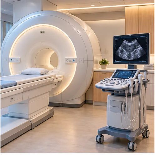

Cramping or pelvic pain is common after embolisation; nausea, tiredness, a short-lived temperature or vaginal discharge may also occur. The clinical team should provide an individualized pain-control plan and clear instructions about follow-up.
Potential complications include infection, unintended injury to blood supply beyond the target, access-site problems, early menopause or changes in ovarian function, persistent or recurrent symptoms, and the need for additional treatment or hysterectomy. Serious complications are uncommon but possible. Ask the treating team about the risks that apply to you.
UAE is not a “one-and-done” guarantee. Hysterectomy remains a possible next step if symptoms persist or return.
This list prioritizes studies of UAE for adenomyosis, newest first. The 2026 evidence review found mostly observational studies with varied definitions and follow-up; the findings cannot predict an individual outcome. This page summarizes the evidence in original plain language.
Studies are provided for discussion with a gynaecologist and interventional radiologist. UAE is generally considered after discussion of established medical and surgical options; fertility goals require individualized counselling.
Contact the practice to ask about appointments or referral requirements.
+91 70348-39446 · veindoctoraju@gmail.com